

LEADERS IN EYE CARE SINCE 1984
WHAT IS GLAUCOMA?

Glaucoma is a group of eye disorders in which the Optic Nerve (the bundle of nerve fibers that carries information from the eye to the brain) gets damaged and can lead to vision loss or blindness. It is also known as ‘ Zamar’.
Optic nerve damage usually occurs in the presence of high eye pressure (high intraocular pressure); but it can also occur with normal or even less than normal eye pressure.
Protect your vision before it is too late! Yes, you read that right Glaucoma is not curable. Although with the help of a Glaucoma specialist in Ahmedabad, early signs of Glaucoma can be diagnosed and treated with the use of medicines,laser or surgery.
Ranchhodrai Eye Hospital is a leading, reputed and trusted eye hospital with the best Glaucoma surgeons in Ahmedabad!
Expert's speak
What Causes Glaucoma?
There are many factors which is contributed in glaucoma.One of major factor is the pressure build up inside the eye. The internal pressure inside the eye is called Intraocular pressure (IOP). The IOP helps maintain and hold the shape of your eye and enables it to function correctly.
So how is the pressure maintained in the eye? Through Aqueous Humor. One part of eyeball(Cilliary processes) produces a clear fluid called the aqueous humor. This fluid is circulated throughout the eye before draining out of your eye. It helps in nourishing different parts of the eyeball. A healthy and normally functioning eye will produce aqueous humor and drain the fluid at the rate at which IOP is maintained within normal range.
Aqueous humor flows out through a mesh-like channel called the trabecular meshwork for draining the fluid. If this drainage channel is clogged or blocked by part of the iris, drainage is hampered thereby pressure builds up inside the eye, which damages the optic nerve.
WHO IS AT RISK FOR GLAUCOMA?
Everyone is at risk for glaucoma.However, certain groups are at higher risk than others.
1) Family History: Presence of glaucoma in other family members especially first degree relative
2) Age: Seen more often in those over 40 years of age
3) Eye Injury: Glaucoma can develop immediately or years after an eye injury
4) Steroids: Patients who are on long term steroid therapy for other diseases such as any ocular disesases or systemic diseases like asthma, arthritis, skin diseases etc.
5) Other Risk Factors are High Myopia (nearsightedness), High Hypermetropia (Farsightedness), Hypertension, Diabetes
SYMPTOMS OF GLAUCOMA
Glaucoma may be asymptomatic
● The signs or symptoms of glaucoma can vary depending on the type of glaucoma.
Primary open angle glaucoma: It develops slowly.The initial symptoms are usually mild and vague and may not be noticed.
● Frequent change of reading glasses.
● Mild eye ache or headache
● Inability to adjust one’s vision on entering a dark room (delayed dark adaptation) Loss of vision, If not treated is first in the peripheral or side vision. The central vision remains intact and is affected last.
Primary angle-closure glaucoma(PACG): In acute attack of PACG,sudden rise of intraocular pressure occurs.
● You experience sudden severe pain in the eye, blurred vision, redness in the eye, colored halos around the light,unilateral headache, nausea, vomiting.
Why Is It Important To Get Regular Eye Checkups For Glaucoma?
-> Glaucoma is also called “the silent thief of vision”, as at first it may not have any symptoms. The first time you notice something is wrong is when significant vision is lost.
-> In older adults, cataract(Motia) usually starts developing at the same age as glaucoma makes an appearance. You may think that you are losing vision due to a cataract when it may actually be due to glaucoma!
-> This is why we advise routine eye check-ups for glaucoma after you turn 40.
-> In the younger population, there are either no symptoms or the onset of symptoms is so slow that it goes unnoticed till there is significant peripheral vision loss.
-> It is important for younger adults, especially those with a family member with glaucoma, to get glaucoma screenings regularly.


How Often Should I Get My Eyes Examined For Glaucoma?
-> Every 1 to 2 years: If you are above 40 years of age or if you have a family member with glaucoma or if you have diabetes or if you are on long term systemic steroids for some other disease, or if you have suffered a blunt eye injury in the past or if you are wearing high plus /high minus number glasses you must get your eyes screened for glaucoma every 1-2 year
-> Every 3 months: If you are diagnosed to have glaucoma Regular review is important and the intervals between review will depend upon the severity of visual loss and the extent of pressure control.
-> Child or a newborn: A child having sensitivity to light or any eye that appears bigger or cloudier than usual, should be shown to an eye specialist to rule out congenital glaucoma.
HOW IS GLAUCOMA TREATED?
● As damage to the optic nerve caused by glaucoma cannot be reversed, the aim of the treatment is to prevent or reduce further damage to the optic nerve.
● The first step is to lower the eye pressure, if high.
● The three main modalities of glaucoma treatment in Ahmedabad are:-
-> Medical (Anti-glaucoma Eye drops and tablets)
->Laser procedure
->Filtering surgery
Medical treatment of Glaucoma
-> Eye drops or tablets are used commonly for early treatment for glaucoma. Medicines lowers IOP either by reducing the production of fluid or by increasing the drainage of fluid out of the eye or by both ways.
-> You must use the medicines timely & regularly as directed by our ophthalmologist.
-> You should not stop your glaucoma medicines without doctor’s advice.
-> You should not buy and use any over the counter medications for Glaucoma.
-> In case if you miss any dose,instill it immediately as soon as you remember without waiting for the next dose.
Laser treatment of Glaucoma
● There are various types of lasers that are used in the treatment of glaucoma. Our glaucoma specialists will decide which laser is suitable for you.
-> YAG Laser Peripheral Iridotomy
-> Selective Laser Trabeculoplasty
-> Argon Laser Trabeculoplasty
->Diode Laser Cycloablation
Filtering Surgery
● Filtering surgery is a kind of “By-pass” surgery of the eye.
● Operation for glaucoma is the only option left for patients in whom the eye pressure is not controlled enough with medication or Laser. It is also the treatment of choice in non-compliant patients & in infants and children with glaucoma.
● Filtering surgery involves creating a drainage opening, to bypass the blockage in the eye’s trabecular meshwork (the eye’s drainage system). This opening helps increase the flow of fluid out of the eye and thereby reduce the eye pressure.
WHY SHOULD I CHOOSE
RANCHHODRAI EYE HOSPITAL FOR GLAUCOMA TREATMENT ?
RANCHHODRAI EYE HOSPITAL FOR GLAUCOMA TREATMENT ?


Is There A Cure For Glaucoma?
As of now, there is no proven or tested cure for glaucoma, but with newer medications and safer surgeries most glaucomas can be stabilized.
We cannot reverse vision loss from glaucoma, however, if detected & treated in time, we are able to control it, slow it down and prevent further vision loss in most people
What Can I Do To Prevent Glaucoma?
Timely detection and treatment is the best way to prevent damage caused by glaucoma.
However, glaucoma itself is not yet preventable. Scientists are researching the triggers of glaucoma and using tests to predict when and who will develop glaucoma.
Until then, regular eye checkups are the only way.
HOW IS GLAUCOMA DIAGNOSED?
Glaucoma is diagnosed after a comprehensive eye check-up and investigations for glaucoma.
When you visit our glaucoma doctor in Ahmedabad we take a detailed patient history and understand your eye problems and do some eye tests for glaucoma.
Here is a summary of the glaucoma tests which may be done during a glaucoma checkup at Ranchhodrai Eye Hospital.
Complete Glaucoma treatment with cutting edge technology
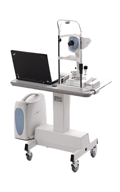
Slit lamp Biomicroscope with 90D lens
The binocular slit-lamp examination provides a stereoscopic magnified view of the eye structures in detail, which helps in diagnosis of Glaucoma. Used with 90 D lens held close to the eye, the slit lamp also provides detailed examination of the Optic disc which gets damaged in Glaucoma.

I Care tonometer
Tonometry by I Care tonometer is a quick,painless and simple test that checks the pressure inside your eyes. The results can help your doctor see if you're at risk for glaucoma. The pressure inside your eye is called intraocular pressure (IOP).

Optical Coherence Tomography(OCT)
The Optovue OCT has good sensitivity and specificity for differentiating normal from glaucomatous eye. It is well known that significant structural nerve fibre loss occurs prior to the development of functional visual field loss.[7] So in this scenario, OCT is especially useful in helping to diagnose glaucoma prior to the onset of visual field loss. OCT also allows for an objective method of visualizing the anterior segment angle(Drainage angle).
Microscope
World best Carl Zeiss Opmi Lumera I Microscope from Germany is the high tech modern optical instrument in this modern era of ophthalmic surgeries. It provides the surgeon with a magnified and illuminated high-quality image of the small ophthalmic structures during high precision glaucoma surgery. Being binocular the surgical microscopes gives the additional benefit of high-quality stereoscopy.

Gonioscopy
Gonioscopy is a eye test which is used to check a part of your eye called the drainage angle. This area is at the front of your eye between the iris and the cornea. It is where fluid called aqueous humor naturally drains out of your eye. Your ophthalmologist will perform a gonioscopy with use of special lenses to check to see if this drainage angle is functioning properly. If the drainage angle is blocked or closed, you may have glaucoma.

Direct ophthalmoscope
Direct ophthalmoscope: Direct ophthalmoscope offers the most magnified view of the optic disc, but the view is not stereoscopic

Indirect ophthalmoscope with 20 D lens
Indirect ophthalmoscope with 20 D lens: the indirect ophthalmoscope delivers a more powerful source of light. It also offers greater scope for stereoscopic inspection of the Optic Disc.

Visual Field Test
Your visual field is how wide of an area your eye can see when you focus on a central point.
Visual field testing is one way your ophthalmologist measures how much vision you have in either eye. For example, if you have glaucoma, this test helps to show any possible side (peripheral) vision loss from this disease

Testimonials

Dr.Bhoomika Jodhani is the best Glaucoma Doctor. I want to share my fantastic experience with Dr. Bhoomika Jodhani, a top-notch glaucoma specialist. She did an amazing job helping my mom with her eye condition. She found the best way to treat her without doing surgery that wasn't needed. She's not only really good at what she does but also cares a lot about her patients. Thanks to her, my mom's eyes are doing much better. I highly recommend Dr. Bhoomika Jodhani for anyone who needs help with their eyes.